Written by Dr Elle Burton-Bradley, Veterinarian, The Village Vet
Lumps and bumps occur in our pets of all ages, however they are more common as our pets get older. We recommend getting all lumps checked by a veterinarian, as they cannot be diagnosed based on sight or location alone.
There are two main categories of lumps: benign and malignant. Most lumps are benign (non-cancerous) and depending on the size and location your veterinarian will determine if it should be removed or if it can be monitored. Some lumps are malignant (cancerous) which may require biopsy or complete removal. We recommend these lumps be sent to the pathologist for confirmatory diagnosis.
Some lumps may be infectious caused by a bacteria, fungus, or virus. Occasionally nipples or ticks can be mistaken for a lump!
So, what happens when our veterinarians check your pet’s lump? During your consultation your veterinarian will examine the lump and will likely stick a needle into it (fine needle aspirate) to look at the cells under the microscope (cytology). In some cases, our veterinarians may want to send the cells to a veterinary pathologist who specialises in identifying cells.
What You Need to Know
Here are some definitions you may want to understand if your Veterinarian finds a lump on your pet:
Fine needle aspirate (FNA): This is when a small needle is inserted into the lump to collect cells which are then sprayed onto a glass slide. The veterinarian will stain this slide and look under the microscope to examine the cells (cytology).
This may be performed in the consultation, or your pet may require a light sedation.
Impression smear: If the lump is ulcerated or has discharging fluid, a glass slide can be pressed against it to collect some cells. The veterinarian will stain this slide and look under the microscope to examine the cells (cytology).
Cytology: This involves staining of the cells on a glass slide and evaluation under the microscope. If your veterinarian is concerned or would like a confirmatory diagnosis, they may recommend sending the samples to a veterinary pathologist for interpretation.
The Village Vets has an onsite digital Imagyst microscope that allows us to scan and digitally send the image of the stained slide to a veterinary pathologist for an efficient and accurate pathology report. This is one component of our goal to reduce our environmental impact and significantly speeds up the test results. In most instances, our clients will receive results during the consult time.
Biopsy: under general anaesthesia a small portion of the lump can be removed (incisional) or the lump can be fully removed (excisional) for pathologist evaluation.
Culture: If the veterinarian is suspicious of an infectious cause, they may send samples to the laboratory for a culture to determine the underlying infectious agent (bacteria, virus, fungus).
Benign (Non-Cancerous) Lumps
Benign lumps are unlikely to spread or invade other areas of the body. They may require removal if they are impeding movement or causing discomfort to your pet.
Some examples include:
- Lipomas: fatty lump, these usually don’t require removal unless they are impeding movement or irritating the pet.
- Abscesses: these are pockets of pus usually due to a penetrating injury (e.g. cat fight wound, stick injury) or a stuck foreign body (e.g. grass seed, deep hair follicle).
- Sebaceous cysts: blocked sebaceous glands.
- Sebaceous adenomas: skin “warts”, these look a bit ugly and usually don’t require removal unless they are irritating the pet or for cosmetic reasons.
- Histiocytomas: these lumps usually occur in young dogs and can appear ulcerated and aggressive and should be examined by your veterinarian. Fortunately, they self-resolve over weeks to months, but may require removal if they are irritating your dog.
- Oral / lip warts: common in puppies and immunocompromised dogs caused by a papillomavirus spread between dogs. These will self-resolve over several months but should be removed if they are bothering the dog or becoming ulcerated.
- Mammary adenomas: benign mammary tumours, these may become malignant over time.
- Meibomian gland tumour: slow growing tumours of the meibomian glands of the eyelids. These are benign tumours but commonly irritate dogs due to their location and readily become inflamed, painful and ulcerated, which may lead to self-trauma of the eye.
Malignant (Cancerous) Lumps
Lumps or tumours that commonly invade and spread to other organs or tissues (metastasis). Early detection and treatment of cancerous lumps increase the chance of a cure for your pet.
- Mast cell tumours (MCT): The most common skin cancer in dogs. Surgical removal of MCTs is necessary. The tumour is sent to a veterinary pathologist to classify them as low or high grade which determines how aggressive they are and the prognosis for your pet. Further specialist treatments may be recommended such as chemotherapy or radiation for high grade tumours.
- Soft tissue sarcoma (fibrosarcoma): Fast growing skin tumours that are locally invasive, look and feel similar to lipomas. As they invade locally wide margins are required for complete removal.
- Melanomas: Tumours of the skin’s pigmented cells, these can be benign or malignant. Those occurring in the mouth or legs often carry a poorer prognosis than those elsewhere on the body. Surgical removal is usually necessary as they readily spread to other organs.
- Squamous cell carcinoma (SCC): Tumours of the skin’s unpigmented cells caused by sun exposure, and commonly appear as crusts or scabs on the nose, lips, eyelids, tips of ears, and vulva. These tumours invade locally and can cause pain and discomfort.
- Mammary carcinomas (breast cancer): cancerous growth of the mammary glands most common in entire (not desexed) female dogs but can occur in cats and male dogs. These cancers spread via the lymph nodes and to other organs.
- Skin (epitheliotropic) lymphoma: rare in our pets, these tumours are known as the “great pretenders” as they can appear as anything from a localised rash to an ulcerated mass.
Common Treatment Options
- Monitor for changes if your veterinarian deems appropriate.
- Surgical removal of part or all of the lump.
- Removal by laser or freezing.
- Chemotherapy.
- Radiation therapy.
Some Concerning Behaviours of Lumps:
- Rapidly growing.
- Ulcerated or bleeding.
- Irritating to the pet.
- Multiple lumps.
- Any change that you have noticed.
- Change in your pet’s behaviour (quiet, flat, reduced appetite).
What You Can Do At Home
It is really helpful for our veterinarians if you keep a record of your pet’s lumps and their changes (e.g. change in size, shape or appearance) over time.
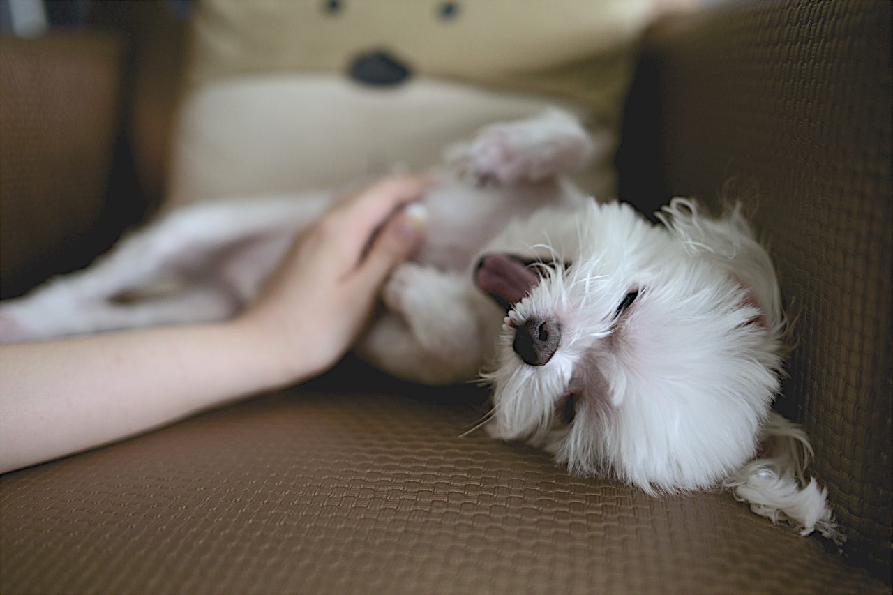
Grooming your pet with a daily brush or cuddle is not only a great bonding experience. Checking your pets regularly with your fingers, feeling through their fur also allows you to find unusual lumps and bumps early on, before they become more serious.
The Village Vet recommend checking your pets regularly and if you notice any new or changing lumps, contact us to schedule a consultation for our veterinarians to thoroughly examine your pet and assess their lumps and bumps!
Book online at The Village Vet or call us directly at the Pymble Clinic on 9499 4010 or Killara Hospital on 8350 5678.
Sources
Dr Elle Burton-Bradley BAnVetBioSci (Hons I) DVM, Veterinarian, The Village Vet


